Mechanism of Muscle Contraction Biology Diagrams
Mechanism of Muscle Contraction Biology Diagrams Learn about the different types of muscle contractions based on force and length, and how they are produced by the sliding filament theory. Find out how muscle tension, length, and force change during isometric, isotonic, concentric, and eccentric contractions. Learn about the process of muscle contraction, the proteins involved, the types of contraction, and the mechanisms proposed to explain it. Find out how actin and myosin filaments slide, how Ca++ ions and ATP are involved, and how nerve impulses trigger muscle contraction.
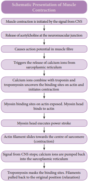
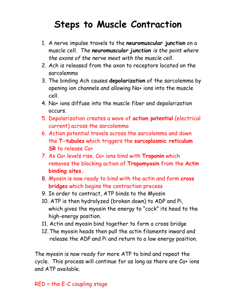
The complex process leading to muscle contraction, called excitation-contraction coupling, begins when an action potential causes depolarization in the myocyte membrane. The depolarization is spread via the transverse (T) tubules, invaginations of the muscle cell membrane, that help spread depolarization signals to the entire muscle fiber. Learn how skeletal muscles contract and relax to move the body, and how the nervous system signals trigger the process. See the 3 steps of muscle contraction, the role of acetylcholine and calcium, and a video explanation.

How Do Muscles Contract: Steps to Muscle Contraction Biology Diagrams
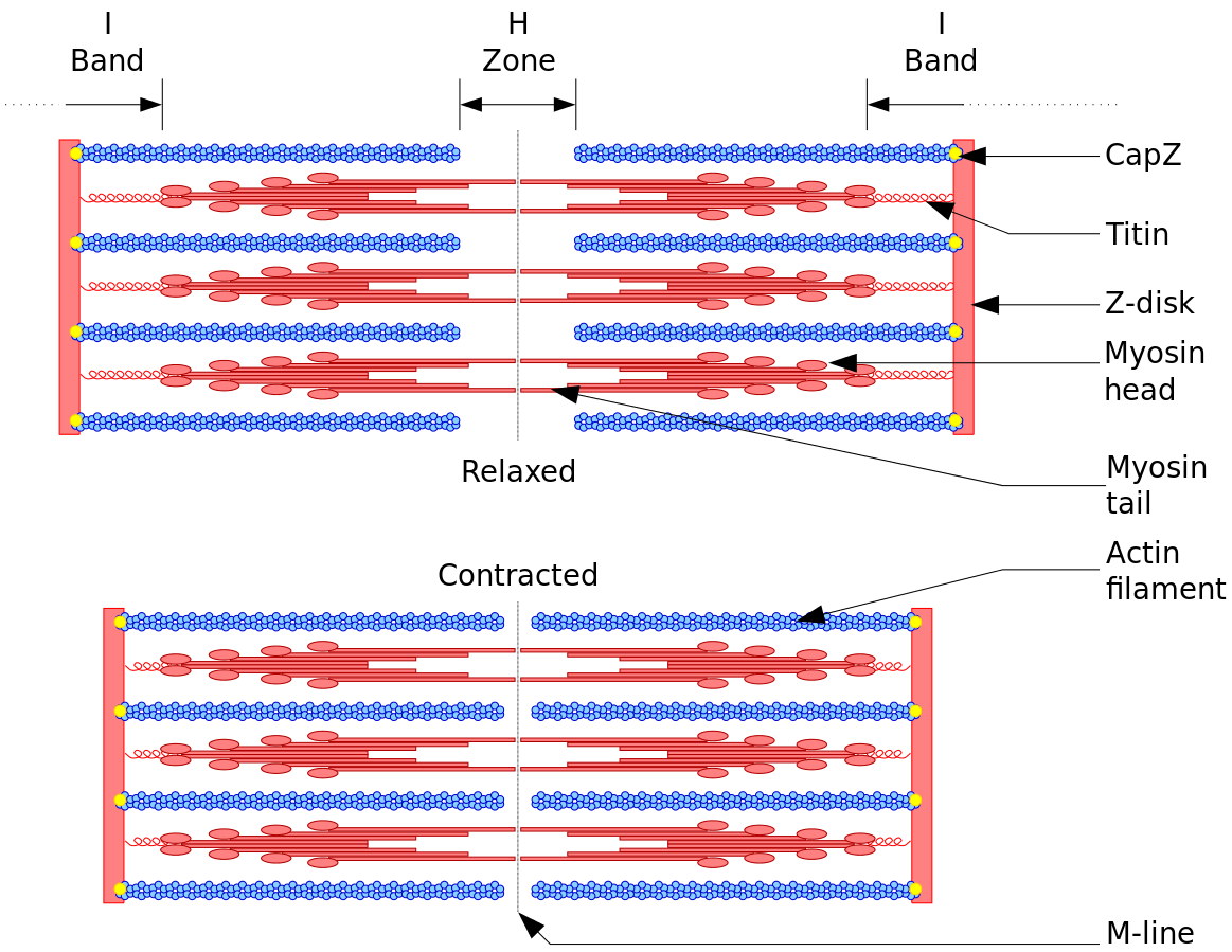
Learn how actin and myosin filaments slide past each other to produce muscle tension and shorten the sarcomere. See diagrams, analogies, and examples of the sliding filament theory and its molecular mechanisms. This entire process shortens the sarcomere, which is functional unit of a muscle cell. You can read a textbook, or even study the simplified steps above, but watching it in action is very helpful. Here's a short snippet of a simulated muscle contraction showing the parts of a muscle cell and what happens during a muscle contraction.
Muscle contraction is the tightening, shortening, or lengthening of muscles when you do some activity. It can happen when you hold or pick up something, or when you stretch or exercise with weights.
Learn Muscular Anatomy - Visible Body Biology Diagrams
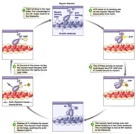
To initiate muscle contraction, tropomyosin has to expose the myosin-binding site on an actin filament to allow cross-bridge formation between the actin and myosin microfilaments. The first step in the process of contraction is for Ca ++ to bind to troponin so that tropomyosin can slide away from the binding sites on the actin strands. This Learn how motor neurons signal skeletal muscle fibers to contract and relax through the neuromuscular junction, excitation-contraction coupling, and calcium handling. Explore the structure and function of the sarcoplasmic reticulum, T-tubules, and cross-bridge cycling.